안녕하세요 오늘은 1일차에 이어 single-cell multiomics를 위한 시퀀싱 기술들을 정리해보려고 합니다.
[대학원 준비 1일차] multiomics란?
안녕하세요, 전에 인체 시스템 정리에 이어 이제 논문을 읽으며 논문 내용을 정리해보려 합니다. 그런 차원에서 오늘 저녁을 기점으로 공부 기록할 수 있는 만큼 기록하면서 일 수를 세보려고
tkmstudy.tistory.com
그러기 전에 본 분석을 수행하기 위해선 동일 세포에서의 multipe types의 분자들을 isolate시키고
해당 분자들을 barcoding하는 절차가 필수1)인 만큼
single-cell mulitiomics analysis를 위한 'cell isolation'와 'barcoding'절차를 소개해보도록 하겠습니다.

참고로 챗GPT가 말하길, 분석 전 위와 같은 절차를 해야하는 이유는
multiomics 분석을 할 수있도록 개별 세포들의 cell-specific context를 보존하고,
cellular heterogeneity의 높은 해상도 view를 가능하게 하기 위함이라는데 맞는지는 논문1)에 적힌 절차들을 정리해보면서 알아보겠습니다.
1. Isolation of single cell
single-cell isolation은 capture methods를 적용하여 dissociated cell suspension으로부터 single cells를 붙잡은 후
mechanical 혹은 enzymatic dissociation을 하는 절차로 이어집니다.

isolation 과정에서 cell을 capturing한다는게 무슨 의미일까해서 chatGPT에게 물어보니까
많은 세포들이 포함된 complex mixture에서 individual cell을 isolating함으로써
획득한 molecular data(gene expression, protein levels)가 특정 세포에 기인할 수 있도록 하여
특정 세포에 대한 rna 시퀀싱이나 multiomics 분석을 가능하게 하는 것이라고 합니다.
무수한 세포에서 나온 transcriptome data가 어떤 세포에서 나온 data인지 특정되지 못한다면,
특정 세포에서 어떤 유전자가 발현되고, 어떤 단백질이 합성되도록 하는지 파악하기 어려울 것이고
결국 몸 속 세포의 발현을 제어함으로써 질병 프로세스를 겨냥하는 치료 전략을 수립하기 어려울 것입니다.
따라서 몸속 무수히 많은 세포로부터 개별 세포를 분리하여 시퀀싱과 multiomics 분석을 진행하는 것입니다.
예로, 해당 분석을 통해 암세포와 정상세포 발달의 차이에서 기인한 genetic heterogeneity를 이해하고 이를 타겟팅할 수 있을 것입니다.
"As the isolation approaches were flourished, the genomics with single cell sequencing technologies opened up new insights to link functionality and phenotype of cell with genotype. Thus, enabling us to know genetic heterogeneity present in individual due to difference in development of normal and diseased cell." - Biocode

논문1)에 따르면, single-cell mono-omics에 쓰는 capture 방법들이 single-cell multiomics에도 적용된다고 하는데
예로, 10~100개 정도의 세포를 붙잡는 low-throughput methods로 laser capture microdissection(LCM)과 robotic micromanipulation이 활용되고,
수천 수만의 세포를 붙잡는 high-throughput methods로는 fluorescence-avtivated cell sorting(FACS)가 활용된다고 합니다.
적은 수의 세포를 붙잡는 방법은 고립시킨 세포의 spatial information(공간적 정보)가 유지된다는 점에서
많은 수의 세포를 붙잡는 방법에 비해 이점이 있기에 각각의 목적에 따라 다른 방법이 활용될 수 있겠습니다.
본 영상에 따르면, LCM은 타깃세포에 Laser에 초점을 두어 해당 세포를 분리한 후(cutting)
다시 defodused하고 그 세포를 transfer vessel에 옮기는 듯 합니다(catapulting).
이 기술은 computer의 도움을 받은 dissection을 사용하여 solid tissues로부터 세포를 분리할 수 있지만, low throughput이라는 한계를 가진다고 합니다.
그러면서도 정확하고, 빠르게 세포의 pure populations을 분리해낼 수 있는 이점이 있다 합니다.
LCM가 만들어내는 산출물에 대해 본 영상을 제작한 'Biocode'에서 글2)로 더 자세하게 소개하고 있습니다.
"LCM results in liquification of thin and transparent thermoplastic film from the cells of interest. The film, which is liquified, then fuse with target cells. Then, as the thermoplastic film is removed which now have the cells of interest. In addition to this, these isolated cells can be centrifuged with appropriate buffer solution for analysis"
즉, LCM을 통해 산출된 관심있는 세포가 있는 liquification 형태의 film을 target cells와 fuse하고,
그럼으로써 필름이 제거되어 관심있는 세포만 분리되었을 때
분석을 위해 적절한 buffer solution과 함께 원심분리를 시킵니다.
이는 본 세포 내용물들(RNA, DNA, 그리고 Protein)의 quality를 유지하기 위함으로
적절한 buffer는 바깥 환경에서 빠르게 degrade되기 쉬운 해당 세포 내용물들을 안정화하여 integrity를 보존합니다.
2. barcoding
이렇게 분리된 세포로부터 DNA, RNA, 단백질 같은 내용물들을 분리하는데
생물학 시간에 배웠듯이 gDNA(genomic DNA)는 핵(nucleus)에 위치하고, mRNAs의 다수는 세포질(cytosol)에 위치합니다.

이때 plasma membrane-selective lysis buffer와 원심분리를 거쳐 세포질로부터 핵을 분리해내게 되면
핵에 있는 gDNA와 세포질에 있는 mRNAs는 잡아낼 수 있지만, 핵에 위치한 mRNAs는 잡아내지 못해 잃어버리게 된다고 합니다.
그 이유는 Plasma membrane-selective lysis buffer는 plasma membrane을 premeabilize 혹은 파열하면서도 nuclear membrane intact를 유지하도록 설계되었기에
해당 buffer를 통한 lysis 과정에서 nucleus에 있는 mRNAs가 방출되지 않아 추출되지 때문입니다.
따라서 mRNAs를 gDNA로부터 분리하여 capture하기 위해서는 'oligo-DT-coated magnetic beads'가 사용됩니다.
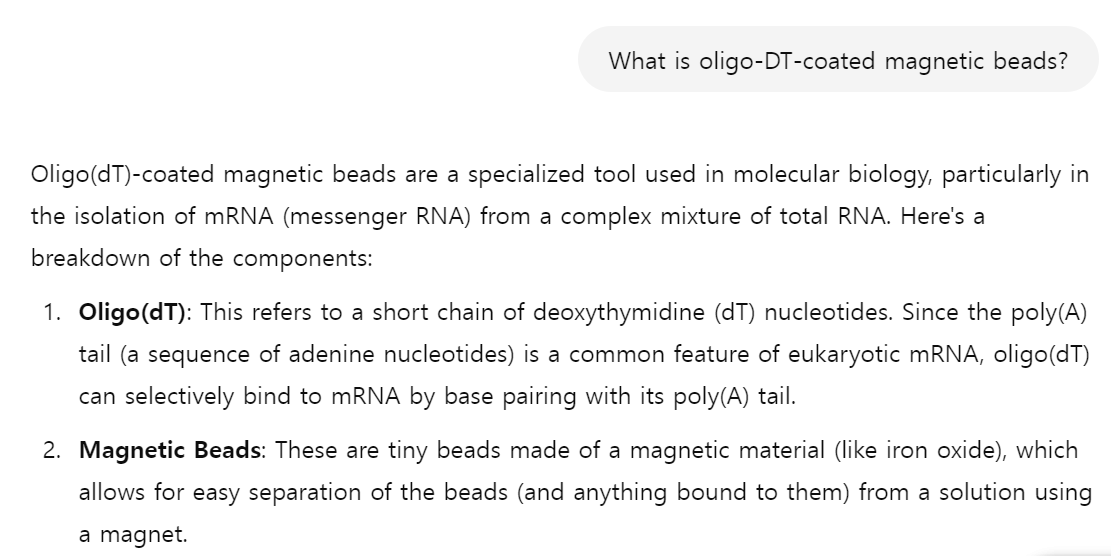
참고로 어제 링크한 영상에서 'Oligo dT'가 등장3)하는데요,

이 Oligo dT는 전사체의 3'에 있는 Poly A tail을 탐지하여 RNA만 따로 뽑는 Poly A detection을 하는데 활용된다고 합니다.
그리고 서로 다른 barcodes(cell-, molecule- identifying barcodes)들이 gDNA와 mRNA의 tract를 구분하여 식별하는데 사용된다1)고 합니다.

이러한 clinical samples는 flash-frozen 혹은 paraffin에 임베드가 되는데 본 freezing 과정은 cytoplasmic membrane은 방해하지만 nuclear membrane은 방해하지 않기에
gDNA와 nuclear mRNA와 달리 cytosolic mRNAs는 single-cell multiomics analysis에서 잘못된 결론을 도출할 수 있다4)고 합니다.
다음 글에선 이제 진짜로 준비된 isolated cells의 mutiple biomolecules(gDNA, mRNAs 등)를 가지고 시퀀싱하는 기술을 소개하도록 하겠습니다.
참고자료
1) Lee, J., Hyeon, D.Y. & Hwang, D. Single-cell multiomics: technologies and data analysis methods. Exp Mol Med 52, 1428–1442 (2020)
2) Biocode, Single Cell Genomics, August 29, 2022, URL : https://www.biocode.org.uk/?s=LCM
LCM | BioCode
As the technique is named as single cell genomics (SCG), so the first step is to isolate single cells for analysis. There are different principles used for cell isolation and on the basis of these we can classify cell isolation techniques into two groups.
www.biocode.org.uk
3) Joyful Bioninfo, RNA-seq 원리 | 전사체, 유전자 발현 분석, Youtube
4) Navin,N. E. Thefirst five years of single-cell cancer genomics and beyond. Genome Res. 25,1499–1507 (2015).

'생물정보학(바이오인포매틱스)' 카테고리의 다른 글
| [대학원 준비 3일차] single-cell RNA sequencing (scRNA-seq)에 대하여 (1) | 2024.09.21 |
|---|---|
| [대학원 준비 2일차] multiple displacement amplification(MDA) & multiple annealing and looping-based amplification cycles (MALBAC) (3) | 2024.09.20 |
| [대학원 준비 1일차] multiomics란? (6) | 2024.09.19 |
| 12. 생식계 :: 정자를 만드는 남성의 생식기관, 난자를 만드는 여성의 생식기관, 그리고 수정 & 착상, 아기 탄생 (2) | 2024.09.18 |
| 11. 내분비계 :: 호르몬을 분비하는 갑상선, 부갑상선, 이자의 랑게르한스섬 등 (3) | 2024.09.18 |


